Molecular Biology
Electrophoresis kits and related items
ELECTROPHORESIS KITS
A classic practical for demonstrating the action of different restriction enzymes on DNA
This innovative practical work uses modern DNA technology to help students learn about classical Mendelian inheritance.

In this module students can amplify chloroplast DNA using the polymerase chain reaction (PCR). The length of the fragments produced can be used to infer evolutionary relationships.

The NCBE’s electrophoresis equipment can be used to analyse proteins as well as DNA. You do, however, need a special type of agarose to carry out this work.
NEW LOWER PRICE!

A crime scene investigation kit, in which the students get to investigate which of the suspects committed the crime by using DNA analysis.
RELATED EQUIPMENT
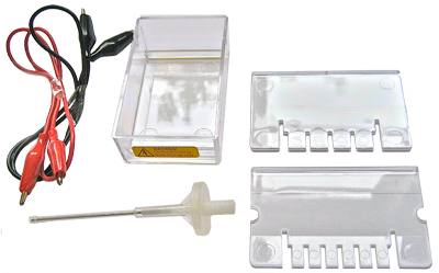
This inexpensive, safe, equipment was developed by the NCBE so that school students could carry out gel electrophoresis with DNA and proteins. It was awarded the coveted Millennium Product status by the Design Council in 2000.
This inexpensive, safe, 36 volt mains transformer allows up to four NCBE gel tanks to be powered simultaneously. Gels typically take two hours to run at this voltage.
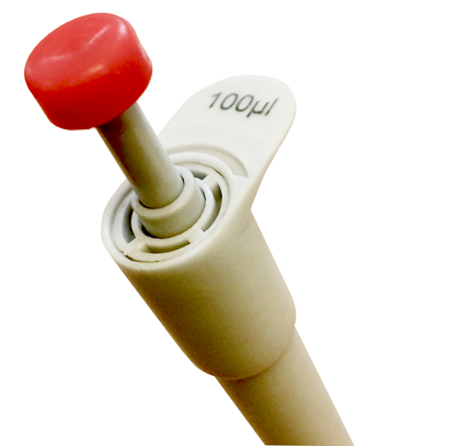
Unlike other cheap pipettes, these ‘Volac MiniPipets’ are accurate, autoclavable and robust. They even have a ‘double action’ like conventional micropipettes.
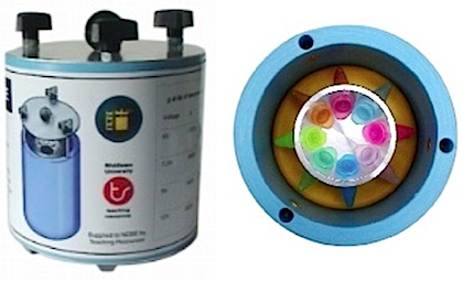
This award-winning 12 volt microcentrifuge holds eight standard (1.5 mL) microcentrifuge tubes, and, unlike many small microcentrifuges, will spin at up to 13,000 rpm (8,500 g), which is sufficient to spin down plasmid DNA, for example.
OTHER ITEMS
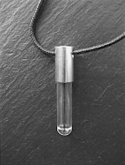
This kit provides sufficient materials to make 30 attractive pendants containing DNA extracted from human cheek cells. It is ideal for science demonstrations on open days, feeder school visits, pupil incentives or fund-raising events such as school fetes.
ABOUT GEL ELECTROPHORESIS
Gel electrophoresis is a key technique in modern biology that features in all the new A Level Biology specifications in England. It is a way of separating DNA, RNA or proteins based on their size and the electrical charge on the molecules.
How it works
First, a gel is cast from agarose; a very pure form of agar, which is obtained from seaweed. At one end of the slab of gel are several small wells, made by the teeth of a comb that was placed in the molten agarose before it set. A buffer solution is poured over the gel, so that it fills the wells and makes contact with the electrodes at each end of the gel. Ions in the buffer solution conduct electricity. The test samples (DNA fragments) are mixed with a small volume of loading dye. This dye is dissolved in a dense sugar solution, so that when it is added to the wells, it sinks to the bottom, taking the sample with it. An electrical potential is applied across the gel. Phosphate groups give the DNA fragments a negative electrical charge, so that the DNA migrates through the gel towards the positive electrode. Small DNA fragments or proteins move quickly through the porous gel; larger molecules travel more slowly. In this way the pieces of DNA or proteins are separated by size. The loading dye also moves through the gel, so that the progress of the electrophoresis can be seen (the DNA itself is invisible).
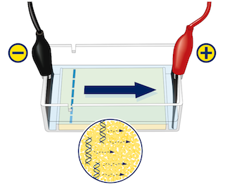
Visualising DNA
After electrophoresis, the DNA is visualised. In research laboratories, a fluorescent dye will have been incorporated into the agarose gel before it was cast. After the gel has been ‘run’ it is illuminated with ultraviolet (UV) light and the dye, which binds to DNA, will show up as bright fluorescent bands. Ethidium bromide is a dye that until recently was most commonly used for staining DNA. Ethidium bromide and its breakdown products are thought to be potent mutagens and carcinogens and therefore it should not be used in schools. Ethidium bromide has similar dimensions to a base pair in DNA. When ethidium bromide binds to DNA, it slips between adjacent base pairs and stretches the double helix. This explains the dye’s mutagenic effect — the ‘extra bases’ cause errors (frameshift mutations) when the DNA replicates. In addition, short-wavelength UV light (which itself is harmful) is required for ethidium bromide to fluoresce and reveal the DNA. For reasons of safety and because UV light of this wavelength causes unwanted mutations in the DNA being studied, several alternative stains are now commonly used in labs. These include SYBRsafe and GelRed, which although they are thought to be safer than ethidium bromide, are far more expensive [Ethidium bromide costs £4.50 per mL compared with £133 per mL for SYBRsafe and £200 per mL for GelRed (2016 prices).]
In schools, safer, cheaper dye solutions are used to stain the entire gel after electrophoresis. Suitable stains include Azure A and Azure B, Toluidine blue O and Nile blue sulphate. This type of stain is not thought to intercalate within the DNA double helix, but instead binds ionically to the negatively-charged phosphate groups of the DNA.
Polyacrylamide gels and protein electrophoresis
If you wish to separate very small DNA molecules or proteins, gels cast from polyacrylamide are sometimes used. Note: For safety reasons, polyacrylamide gels should not be cast in schools: the acrylamide use to make the gels is a neurotoxin. (You can use pre-cast gels if you wish, but they have a limited shelf life.)
It is possible to separate proteins using a special type of agarose, but in contrast to the procedure using polyacryamide, the proteins are separated by charge only (not charge and size). This is because the pores within the agarose gel are relatively large and the proteins can easily pass through them.
As with DNA, the proteins on the gel are stained with an appropriate dye such as Coomassie blue.
UNIQUE, AWARD-WINNING, SAFE, TECHNOLOGY
The enzymes and DNA in the NCBE’s award-winning kits are dried. These reagents can be transported and stored at room temperature instead of the -20 °C that is normally required for such delicate biological molecules. To dispense small volumes of reagents with the required precision, the NCBE has coupled special microsyringes with calibrated tips, providing a low-cost yet highly accurate alternative to conventional micropipettes. The NCBE’s gel electrophoresis apparatus uses carbon fibre electrodes rather than the platinum that is normally used. The equipment is powered by low-voltage batteries or an inexpensive, safe and effective 36 volt mains transformer. To stain the DNA on the gel the kits use Toluidine Blue O in preference to fluorescent stains and ultraviolet light. If a special agarose is used, proteins can also be separated using this equipment and stained with Colloidal Coomassie Blue.
THE POLYMERASE CHAIN REACTION
The polymerase chain reaction (PCR) is one of the most important and powerful methods in molecular biology. It enables millions of copies of specific DNA sequences to be made easily and quickly. The technique and variations of it are used extensively in medicine, in molecular genetics and in pure research. The PCR and plant evolution module provides materials for the simple extraction of chloroplast DNA from plant tissue, its amplification by the PCR, and gel electrophoresis of the PCR product. Students can use plants of their choice and identify possible evolutionary relationships between different species. This mirrors the molecular methods used in modern plant taxonomy. This activity presents an ideal opportunity for open-ended investigations by individual students or groups.